pleural effusion chest x ray
Diagnostic aspiration which may be included in therapeutic aspiration. Chest radiographs are the most commonly used examination to assess for the presence of pleural effusion.

Tuberculous Pleural Effusion Brown Emergency Medicine
This is a common finding on chest X-ray which can have many causes such as.

. Related videosIntroduction to Chest X-ray Interpretation. Pleural effusions are a rare complication of metastatic adenocarcinoma of the prostate and have not been noted to resolve with endocrine therapy. Possible causes include Bronchogenic Carcinoma.
Check the full list of possible causes and conditions now. A unilateral effusion is typically exudative whereas. Chest X-Ray Abnormal Hoarseness Pleural Effusion Symptom Checker.
Check the full list of possible causes and conditions now. You may find out you have pleural effusion through a chest X-ray or physical examination done for another reason. Possible causes include Renal Cell Carcinoma.
A pleural effusion is a collection of fluid in the pleural space. Visceral pleura and parietal pleura that. Pleural Effusion on Chest X-Ray Motor Neuron Disease Symptom Checker.
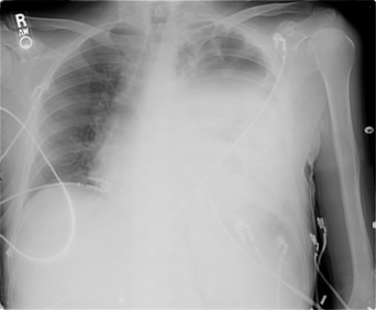
Asymmetric pleural effusions Pleural effusions caused by heart failure may not be symmetrical This patient with heart failure had been nursed lying on their right side before this X-ray was. Pleural effusion can be diagnosed through physical examination chest X-ray CT scan and ultrasound. Chest x-ray Computed tomography CT scan of the chest Ultrasound of the chest Thoracentesis a needle.
A lateral decubitus projection is most sensitive able to identify even a sm See more. A prediction rule was. In a patient with massive bilateral pleural.
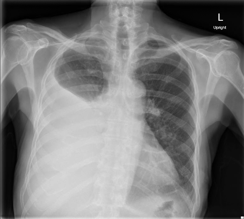
X-ray Frontal Obliteration of left costophrenic angle with a wide pleural based dome shaped opacity projecting into the lung noted tracking along the CP angle and lateral chest wall. This is useful to assess a pleural effusion and estimate its size. The tests most commonly used to diagnose and evaluate pleural effusion include.
However it should be noted that on a routine erect chest x-ray as much as 250-600 mL of fluid is required before it becomes evident 6. Furthermore the treatment options for pleural effusion include. If the patient is upright when the X-ray is taken.
A pleural effusion is the accumulation of fluid between the layers of pleura that cover the lung. A chest X-ray is the first-line imaging investigation of choice. 12 Normally the space between the visceral pleura and the parietal pleura cannot be seen.
The first step in diagnosis is confirmation of the pleural effusion suspected clinically or on chest x-ray by ultrasound. When a doctor examines you they may notice expansion. Fluid gathers in the lowest part of the chest according to the patients position.
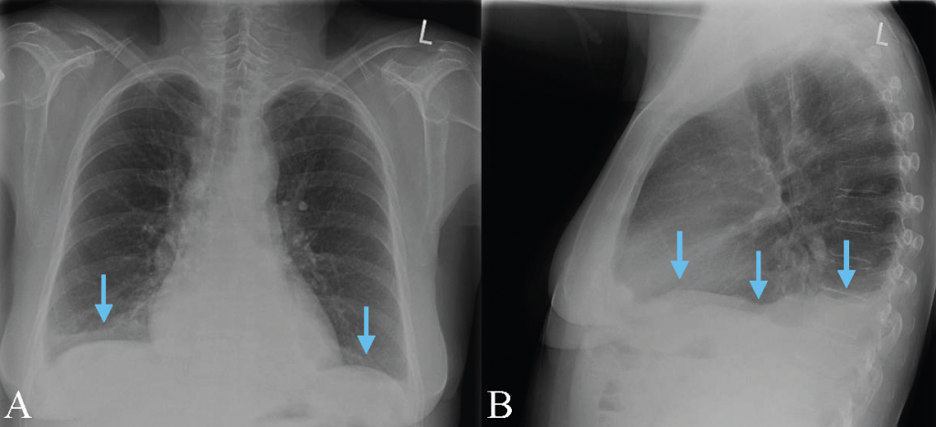
We devised a prediction rule for estimating pleural effusion volume on the basis of posteroanterior and lateral chest radiographs. A pleural effusion appears as an area of whiteness on a standard posteroanterior chest X-ray. Pleura is a mesothelial lined sac that envelopes the lungs and comprises of 2 membranous walls ie.
Chest X-ray Pleural Effusion.
Case 8 Answers Pleural Effusions Clinical Respiratory Diseases Critical Care Medicine Seattle Med 610 University Of Washington School Of Medicine
Case 6 Pleural Effusions Clinical Respiratory Diseases Critical Care Medicine Seattle Med 610 University Of Washington School Of Medicine
Cureus Unexplained Pleural Effusion Leads To The Revelation Of A Malignant Mesothelioma A Case Report
Pocus For Pleural Assessment And Intervention Pocus Journal
Large Right Pleural Effusion Jetem
Pleural Effusion In Adults Etiology Diagnosis And Treatment 24 05 2019

Pleural Effusion X Ray Stock Image C001 7291 Science Photo Library

Chest X Ray Showing Right Pleural Effusion Download Scientific Diagram

Pleural Effusion Undergraduate Diagnostic Imaging Fundamentals

A Curriculum Learning Strategy To Enhance The Accuracy Of Classification Of Various Lesions In Chest Pa X Ray Screening For Pulmonary Abnormalities Scientific Reports

Local Pleural Effusion Medical Radiography Medical Knowledge Radiology

Chest Xray Severe Right Pleural Effusion Stock Photo 1432542602 Shutterstock
Cureus Two Patients With Meigs Syndrome And Elevated Serum Ca 125 A Case Report

Lti 01 Lung Therapeutics

A Chest X Ray Shows Bilateral Pleural Effusion And Diffuse Interstitial Download Scientific Diagram

Chest X Ray Showing The Massive Pleural Effusion On The Right Side That Download Scientific Diagram
Pleural Effusion Radiology Key

Icu Chest Films

Chest Radiology

Comments
Post a Comment